 |
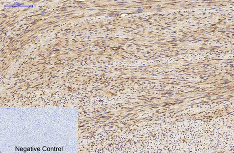
Immunohistochemical analysis of paraffin-embedded Human-uterus tissue. 1,LC3A Mouse Monoclonal Antibody(5G10) was diluted at 1:200(4°C,overnight). 2, Sodium citrate pH 6.0 was used for antibody retrieval(>98°C,20min). 3,Secondary antibody was diluted at 1:200(room tempeRature, 30min). Negative control was used by secondary antibody only. |
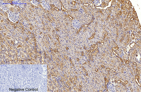 |
Immunohistochemical analysis of paraffin-embedded Rat-kidney tissue. 1,LC3A Mouse Monoclonal Antibody(5G10) was diluted at 1:200(4°C,overnight). 2, Sodium citrate pH 6.0 was used for antibody retrieval(>98°C,20min). 3,Secondary antibody was diluted at 1:200(room tempeRature, 30min). Negative control was used by secondary antibody only. |
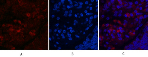 |
Immunofluorescence analysis of Human-lung-cancer tissue. 1,LC3A Mouse Monoclonal Antibody(5G10)(red) was diluted at 1:200(4°C,overnight). 2, Cy3 labled Secondary antibody was diluted at 1:300(room temperature, 50min).3, Picture B: DAPI(blue) 10min. Picture A:Target. Picture B: DAPI. Picture C: merge of A+B |
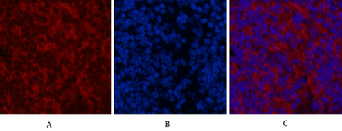 |
Immunofluorescence analysis of Mouse-spleen tissue. 1,LC3A Mouse Monoclonal Antibody(5G10)(red) was diluted at 1:200(4°C,overnight). 2, Cy3 labled Secondary antibody was diluted at 1:300(room temperature, 50min).3, Picture B: DAPI(blue) 10min. Picture A:Target. Picture B: DAPI. Picture C: merge of A+B |
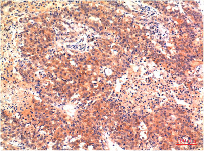 |
Immunohistochemical analysis of paraffin-embedded Human Heptacarcinoma Tissue using LC3A Mouse mAb diluted at 1:200. |
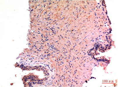 |
Immunohistochemical analysis of paraffin-embedded Human Prostate Carcinoma Tissue using LC3A Mouse mAb diluted at 1:200. |
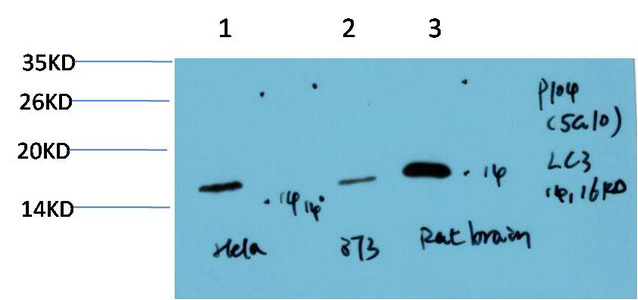 |
Western blot analysis of 1) Hela Cell Lysate, 2) 3T3 Cell Lysate, 3) Rat Brain Tissue Lysate using LC3A Mouse mAb diluted at 1:1000. |

 查看与下载
查看与下载